




Modern prostate MRI has transformed the way suspected prostate cancer is investigated. In some cases, a reassuring MRI result combined with other low-risk findings may mean you do not need an immediate biopsy. However, many men still require tissue sampling to confirm whether cancer is present.
An MRI provides detailed images of the prostate and can help identify areas that may appear suspicious. This information allows doctors to assess your risk more accurately and decide whether a biopsy is necessary. If a biopsy is recommended, the MRI can also help target the most relevant areas for sampling.
Although MRI is a highly valuable diagnostic tool, it cannot always confirm the exact nature of prostate cells. A biopsy remains the only way to examine prostate tissue under a microscope and provide a definitive diagnosis. As a result, MRI has not replaced biopsy entirely, but it has helped make the diagnostic process more selective, targeted, and effective for you.
Understanding What a Prostate MRI Does
A prostate MRI uses magnetic fields and radio waves to create detailed images of your prostate and the surrounding tissues. The scan can identify areas that appear different from normal prostate tissue and may require further assessment. This gives your specialist a clearer picture of what is happening inside the prostate.
MRI is particularly useful because a PSA blood test cannot show where a suspicious area is located. By providing detailed anatomical information, the scan can help your specialist decide whether additional investigations are necessary. This may help you avoid unnecessary procedures in some situations.
Although MRI provides valuable information, it does not examine prostate cells directly. The findings are based on how tissue appears on imaging rather than what the cells look like under a microscope. For this reason, you may still need a biopsy if there is concern about the presence of prostate cancer.
Understanding What a Prostate Biopsy Does
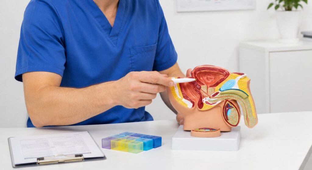
A prostate biopsy involves taking small tissue samples from your prostate gland for laboratory analysis. These samples are examined under a microscope to determine whether cancer cells are present. This makes a biopsy one of the most important tools for confirming a diagnosis of prostate cancer.
If cancer is detected, the biopsy can provide additional information about how abnormal the cells appear. This helps doctors assess the aggressiveness of the cancer and plan the most appropriate treatment for you. The results play a key role in guiding further management decisions.
This is why a biopsy remains important even when MRI has been performed. While an MRI can identify areas that may look suspicious, it cannot confirm exactly what those areas contain. A biopsy provides the tissue-based evidence needed to establish a definitive diagnosis and guide your care.
Why MRI Is Often Performed Before Biopsy
Modern diagnostic pathways often use a prostate MRI before biopsy when prostate cancer is suspected. This approach helps your specialist gather more information before deciding whether tissue sampling is necessary. As a result, you may receive a more personalised assessment based on your individual level of risk.
MRI can identify areas within the prostate that appear suspicious and may require closer investigation. It can also help determine whether a biopsy is likely to provide useful diagnostic information for you. If a biopsy is needed, the MRI can guide sampling towards the most relevant areas.
This differs from earlier diagnostic pathways in which biopsy decisions were often made without the benefit of pre-biopsy MRI. Today, your specialist can combine MRI findings with PSA levels, symptoms, and other risk factors to make a more informed decision. This helps ensure that investigations are targeted and appropriate for your situation.
Evidence Note:
The landmark PROMIS study helped establish MRI as an important test before prostate biopsy. In 576 men who completed the study pathway, multiparametric MRI had a sensitivity of 93% for clinically significant prostate cancer under the study definition, compared with 48% for standard TRUS biopsy. The study demonstrated why MRI is useful as a triage test before biopsy, but also showed that MRI cannot provide perfect exclusion of clinically significant disease.
For you, the practical message is that MRI can help identify who is more likely to benefit from biopsy and where suspicious tissue should be sampled. However, it should be used as part of a risk-based pathway rather than viewed as a guaranteed replacement for tissue diagnosis.
Can a Reassuring MRI Help You Avoid Biopsy?
Yes, in some cases, a reassuring MRI may mean that you do not need an immediate prostate biopsy. This decision depends on how reassuring the scan findings are and whether your overall risk of prostate cancer is considered low. Your specialist will assess the MRI results alongside other clinical information before making a recommendation.
Pre-biopsy MRI pathways can reduce the number of biopsies performed by allowing some carefully selected men with reassuring imaging and a low overall clinical risk to undergo monitoring instead of immediate biopsy.
However, avoiding a biopsy does not necessarily mean that no further follow-up will be required. You may still need regular PSA testing, repeat assessments, or additional imaging over time. If your PSA level rises or new concerns develop, your specialist may recommend further investigation, including a biopsy if appropriate.
When Might MRI Reduce the Need for Biopsy?
| Clinical situation | Can biopsy potentially be deferred? | What usually influences the decision? |
| Reassuring MRI and low overall clinical risk | Possibly | PSA, PSA density, PSA trend, examination findings and family history |
| Reassuring MRI but persistently concerning PSA profile | Not necessarily | Further assessment or biopsy may still be recommended |
| Equivocal MRI finding | Depends on overall risk | PSA density, previous biopsy history and other clinical factors |
| Clearly suspicious MRI lesion | Biopsy is commonly recommended | MRI findings guide targeted tissue sampling |
| Suspicious MRI but previous negative biopsy | Further assessment may still be needed | Quality of targeting, PSA trend and persistence of the lesion |
| Previous negative biopsy but ongoing clinical concern | MRI can guide the next step | Repeat imaging, targeted biopsy or further monitoring may be considered |
Important: The decision should not be based on MRI alone. NICE recommends considering MRI findings together with PSA, digital rectal examination findings, comorbidities, risk factors and previous biopsy history.
Why a Negative MRI Is Not an Absolute Guarantee
A reassuring MRI can be very encouraging, but it cannot guarantee that no prostate cancer is present. While MRI is highly effective at identifying many significant cancers, some abnormalities may not be visible on imaging. This means that a normal or low-suspicion MRI does not completely rule out the possibility of cancer.
Your specialist will not rely on MRI findings alone when assessing your risk. Other factors, such as your PSA level, PSA trend, prostate examination findings, age, family history, and overall clinical picture, are also taken into account. Looking at all of this information together helps create a more accurate assessment of your situation.
If your overall risk remains concerning despite a reassuring MRI, a biopsy may still be recommended. The decision is based on more than just how the scan appears. Your specialist will consider whether all available evidence supports monitoring safely or whether further investigation is needed.
Clinical Insight: Negative MRI Does Not Mean Zero Risk
A reassuring MRI substantially reduces concern, but it cannot rule out clinically significant prostate cancer with complete certainty. Published meta-analysis data show that the reliability of a negative MRI varies according to the definition of significant cancer, the patient population, MRI quality and reporting expertise.
Your specialist should therefore consider MRI findings alongside PSA density, PSA trend, examination findings, family history and previous investigations before deciding whether monitoring or biopsy is more appropriate.
What Happens When MRI Finds a Suspicious Area?
If your MRI identifies a suspicious area within the prostate, your specialist may recommend a biopsy for further investigation. The MRI helps show exactly where the abnormal area is located, allowing tissue samples to be taken more accurately. This targeted approach can improve the chances of obtaining useful diagnostic information.
A suspicious finding on MRI does not automatically mean that cancer is present. In some cases, benign conditions, inflammation, or other non-cancerous changes can produce similar appearances on imaging. This is why a biopsy may still be needed to determine exactly what the abnormal area contains.
Your specialist should explain the MRI findings and discuss why a biopsy is being considered. You should have a clear understanding of the level of concern, where the suspicious area is located, and how the biopsy may help provide a definitive diagnosis. This information can help you make informed decisions about the next steps in your care.
MRI-Targeted Biopsy Explained

An MRI-targeted biopsy is designed to sample areas of the prostate that appear suspicious on MRI imaging. The information from your scan is used to guide the biopsy towards these specific locations rather than relying solely on random tissue sampling. This helps make the investigation more focused and precise.
By targeting suspicious areas directly, the biopsy may improve the detection of clinically significant prostate cancer. In many cases, tissue samples are taken from the area of concern as well as from other parts of the prostate when appropriate. This provides your specialist with a more complete assessment of your prostate health.
An MRI-targeted biopsy does not necessarily mean that only a single sample is taken. The number and location of samples depend on your MRI findings, prostate anatomy, biopsy technique, and overall clinical situation. Your specialist will determine the most suitable sampling approach based on your individual needs.
Research Insight:
The PRECISION trial compared an MRI-based diagnostic pathway with standard biopsy in men who had not previously undergone prostate biopsy. In the MRI pathway, 28% of men had MRI results that were not considered suspicious and therefore avoided biopsy. Clinically significant cancer was detected in 38% of men assigned to the MRI pathway compared with 26% in the standard biopsy group.
For patients, this shows that MRI can reduce some unnecessary biopsies while helping direct tissue sampling towards areas more likely to contain clinically significant cancer. However, the findings do not mean that every patient with a negative MRI can safely avoid biopsy.
Why Systematic Samples May Still Be Taken
Although MRI is very useful for identifying suspicious areas, not all prostate cancers are clearly visible on imaging. For this reason, your specialist may recommend taking additional systematic samples from other parts of the prostate during the biopsy. This helps ensure that areas not highlighted on MRI are also assessed.
Combining targeted and systematic sampling can provide a more comprehensive evaluation of your prostate. While targeted samples focus on areas that appear abnormal, systematic samples help reduce the risk of missing cancers elsewhere in the gland. This approach may improve the overall accuracy of the investigation.
Before the procedure, your specialist should explain the biopsy method that is planned for you. You should understand whether the biopsy will involve targeted sampling, systematic sampling, or a combination of both. Having this information can help you feel more informed and prepared for the procedure.
How PI-RADS Scores Influence the Decision
After your prostate MRI, any suspicious areas may be assigned a PI-RADS score ranging from 1 to 5. Lower scores generally indicate a lower likelihood of clinically significant prostate cancer, while higher scores suggest a greater level of concern. This scoring system helps your specialist assess how suspicious a particular area appears on imaging.
A higher PI-RADS score will often strengthen the case for a biopsy, but it is not the only factor considered. Your specialist will also review information such as your PSA level, PSA density, examination findings, and previous biopsy history. Looking at all these factors together helps create a more accurate assessment of your risk.
If your MRI findings are borderline or uncertain, the next steps may require a more individualised discussion. Your specialist may consider whether immediate biopsy or ongoing monitoring is the most appropriate option for you. The goal is to balance the need for accurate diagnosis with avoiding unnecessary procedures whenever possible.
UK Practice Note:
PI-RADS and Likert scoring both use five categories to communicate increasing levels of MRI suspicion, but they are separate systems. NICE specifically frames the UK diagnostic pathway around Likert scoring, while PI-RADS is a structured international prostate MRI reporting system. Your specialist should interpret the score according to the reporting method used rather than assuming the two systems are technically interchangeable.
What Does a Low-Suspicion MRI Mean?
A low-suspicion MRI is generally a reassuring result because it has not identified features that strongly suggest clinically significant prostate cancer. This may reduce the likelihood that an immediate biopsy is needed. However, it is important to remember that a reassuring MRI cannot completely rule out the presence of cancer.
If the rest of your assessment is also considered low risk, your specialist may recommend PSA monitoring rather than immediate further investigation. This approach allows your prostate health to be reviewed over time while avoiding unnecessary procedures. You may still require additional tests if new concerns arise in the future.
Regular follow-up is an important part of this process. You should understand when your next PSA test is due and what changes might prompt further assessment. Having a clear monitoring plan helps ensure that your prostate health continues to be reviewed appropriately.
UK Guidance Note:
NICE recommends multiparametric MRI as the first-line investigation for suspected clinically localised prostate cancer and uses a five-point Likert scale within its diagnostic pathway. MRI-influenced biopsy is recommended for a Likert score of 3 or more. For a Likert score of 1 or 2, biopsy may be omitted after discussing the risks and benefits and reaching a shared decision.
For men with raised PSA and a Likert 1 or 2 MRI who have not undergone biopsy, NICE recommends repeat PSA testing after 3 to 6 months. Biopsy should still be considered where suspicion remains strong, including situations involving raised PSA density, rapid PSA change or strong family history.
What Does an Equivocal MRI Mean?
Sometimes an MRI result is neither clearly reassuring nor strongly suspicious for prostate cancer. You may hear this described as an equivocal or indeterminate finding. In these situations, the scan has not provided a definite answer about whether significant cancer is present.
When this occurs, your specialist will usually consider other factors alongside the MRI findings. These may include your PSA level, PSA density, PSA trend, prostate size, family history, and any previous biopsy results. Looking at the wider clinical picture helps build a more accurate assessment of your risk.
Some men with an equivocal MRI may be advised to have a biopsy, while others may be suitable for monitoring and follow-up. The most appropriate approach depends on your overall risk profile rather than the MRI result alone. Your specialist will discuss the available options and help you understand the reasoning behind the recommendation.
Why PSA Density Can Matter
PSA density compares your PSA level with the size of your prostate. This measurement can provide additional context because a larger prostate may naturally produce more PSA, even when cancer is not present. As a result, PSA density can help your specialist assess your risk more accurately.
MRI plays an important role by providing detailed information about the volume of your prostate. This allows PSA density to be calculated and considered alongside your MRI findings and other clinical factors. Together, these pieces of information help create a more complete picture of your prostate health.
PSA density can be particularly helpful when your MRI result is not clearly reassuring or strongly suspicious. In these situations, it may help determine whether a biopsy or continued monitoring is more appropriate for you. Your specialist can explain how your PSA density influences the decision-making process in your specific case.
Clinical Tip: PSA Density Is Useful, but It Is Not a Standalone Answer
PSA density can add valuable information when MRI findings are negative or equivocal, but it should not automatically determine whether you have a biopsy. Research has shown that combining MRI findings with additional clinical factors can improve biopsy selection compared with using one measurement alone. Your PSA trend, age, examination findings, family history, previous biopsy results and MRI findings should all be considered together.
Why PSA Trend Still Matters After MRI
Your PSA history remains an important part of your prostate assessment, even after you have had an MRI scan. A PSA level that remains stable over time may be viewed differently from one that continues to rise. Looking at the overall trend can provide valuable information about your prostate health.
MRI provides a detailed picture of your prostate at a specific moment, while repeated PSA tests show how things may be changing over time. Together, these findings help your specialist build a more complete understanding of your individual risk. This information can guide decisions about monitoring, further imaging, or additional investigations.
For this reason, it is important that you continue attending any planned PSA tests after your MRI. Even if your scan was reassuring, future changes in PSA levels may influence what happens next. Your specialist may then consider whether repeat imaging or a biopsy is appropriate based on your updated results.
Can MRI Diagnose the Grade of Prostate Cancer?
MRI can identify areas of the prostate that appear suspicious, but it cannot directly determine the grade of prostate cancer. The scan provides detailed images that help assess the likelihood of significant disease, but it does not examine cancer cells themselves. For this reason, MRI alone cannot provide the same level of information as a biopsy.
If cancer is found during a biopsy, the tissue samples are examined under a microscope by a pathologist. This analysis helps determine how abnormal the cancer cells appear and provides important information about the behaviour of the disease. These findings play a key role in assessing the severity of the cancer.
The biopsy results are then considered alongside your PSA level, MRI findings, clinical stage, and other relevant factors. This combined information helps your specialist recommend the most appropriate treatment options for you. While MRI is an important diagnostic tool, pathology remains essential for understanding the characteristics of prostate cancer in greater detail.
Can MRI Confirm That a Suspicious Area Is Cancer?
MRI is very effective at identifying areas that may look suspicious, but it cannot always confirm whether those areas are cancerous. The scan assesses patterns and characteristics within the prostate, but some non-cancerous conditions can produce similar appearances. This means that a suspicious MRI finding does not automatically mean cancer is present.
A biopsy provides a different type of information by allowing tissue samples to be examined under a microscope. This helps determine whether cancer cells are actually present and provides a definitive diagnosis. For many patients, this remains an essential step in the diagnostic process.
Understanding this distinction can help you see why a biopsy may still be recommended after a concerning MRI result. The purpose is not to repeat the same investigation, but to answer a different question about what the suspicious area contains. By combining imaging and tissue analysis, your specialist can make more informed decisions about your care.
Can MRI Find Every Prostate Cancer?
MRI is a highly valuable tool for investigating suspected prostate cancer, but it cannot detect every cancer. Some prostate cancers may not be clearly visible on imaging, which means a reassuring MRI does not completely rule out the presence of disease. This is one reason why additional tests may still be recommended in certain situations.
Despite this limitation, MRI remains extremely useful in the diagnostic process. It can help assess your risk more accurately, reduce the need for unnecessary biopsies, and identify areas that may require targeted sampling. This allows investigations to be more focused and personalised.
The most effective approach is to consider MRI as part of a wider assessment rather than a standalone test. Your MRI findings, PSA results, clinical history, and biopsy information, when needed, all contribute to the overall picture. By combining these sources of information, your specialist can make more informed decisions about your care.
What If Your MRI and PSA Results Do Not Match?

Sometimes your MRI and PSA results may appear to tell different stories. For example, you might have a reassuring MRI scan but a PSA level that continues to rise or remains concerning. When this happens, your specialist will look more closely at all the available information rather than relying on a single test result.
Additional factors such as PSA density, previous PSA trends, clinical findings, and the quality of the MRI interpretation may be reviewed. These details can help determine whether further investigation is needed or whether monitoring remains appropriate for you. A mismatch between results does not automatically mean there is a serious problem, but it does require careful assessment.
In some cases, your situation may be discussed by a multidisciplinary team of specialists. This allows experts from different fields to review your results together and consider the most appropriate next steps. Such collaborative decision-making can help ensure that you receive a thorough and well-informed assessment.
What If Your MRI Looks Suspicious but the Biopsy Is Negative?
A suspicious MRI followed by a negative biopsy can be confusing and may leave you with unanswered questions. However, this does not automatically mean that either test was incorrect. Sometimes the biopsy may not have sampled the exact area of concern, or the MRI finding may be related to a non-cancerous condition.
Your specialist will review the results carefully and consider whether the suspicious area was adequately assessed. Depending on your situation, you may be advised to have repeat PSA testing, further MRI evaluation, additional imaging, or, in some cases, another biopsy. The decision will be based on your overall level of risk and the degree of concern raised by the MRI findings.
The most appropriate next step will vary from person to person. Some patients may only require ongoing monitoring, while others may benefit from further investigation. Your specialist should explain the findings clearly so that you understand why a particular approach is being recommended for you.
Can MRI Replace Repeat Biopsy?
MRI can be very helpful when deciding whether you need another prostate biopsy, especially if you have previously had a negative biopsy but concerns remain. The scan may identify areas that were not sampled adequately during the earlier procedure. This can help your specialist decide whether further investigation is necessary.
However, MRI does not automatically eliminate the need for a repeat biopsy. If a suspicious area remains visible on imaging or your overall risk continues to be concerning, additional tissue sampling may still be recommended. The goal is to ensure that any significant prostate cancer is not missed.
When discussing the next steps, your specialist will consider your previous biopsy results, MRI findings, PSA changes, and other risk factors. These details help determine whether monitoring or another biopsy is the most appropriate option for you. Understanding the reasons behind the recommendation can help you feel more confident about your care plan.
MRI Before a First Biopsy
Having an MRI before your first prostate biopsy is now an important step if localised prostate cancer is suspected. The scan gives your specialist detailed images of your prostate, helping decide whether a biopsy is needed and guiding decisions from the very start.
- Deciding on a Biopsy: MRI can show whether a biopsy is appropriate for you, potentially avoiding unnecessary procedures.
- Targeting Suspicious Areas: If a biopsy is required, the MRI helps identify areas to sample, improving accuracy and reliability.
- Moving Away from Automatic Biopsies: This approach reduces the need to perform a biopsy after every concerning PSA result, making assessment more personalised.
- Understanding Your Pathway: Your individual plan may differ depending on your clinical situation and available services, and your specialist should explain the next steps clearly.
Overall, an MRI before your first biopsy helps you and your specialist make more informed decisions. Knowing the purpose and benefits of each test can help you feel prepared and confident throughout your assessment.
Understanding the Risks of Biopsy
A prostate biopsy is an invasive procedure, which is why avoiding an unnecessary biopsy can be beneficial for you. Possible side effects may include bleeding, discomfort, difficulty passing urine, and infection, although the exact risks vary depending on the biopsy method and your individual circumstances. Your specialist will discuss these potential risks before the procedure.
This is one reason why MRI has become such an important part of the diagnostic pathway. By providing detailed information about your prostate, the scan can help determine whether a biopsy is necessary and identify the areas that may require investigation. This can make the process more selective and targeted.
However, avoiding a biopsy is only appropriate when it can be done safely. If your overall risk of clinically significant prostate cancer remains concerning, the benefits of obtaining tissue samples may outweigh the potential drawbacks of the procedure. Your specialist will help you understand the balance of risks and benefits in your specific situation.
Transperineal and Transrectal Biopsy
Prostate biopsies can be performed using different techniques, and the most suitable option for you will depend on your clinical situation and local practice. The two main approaches are transperineal biopsy and transrectal biopsy. Both methods are designed to obtain tissue samples that can be examined for signs of prostate cancer.
A transperineal biopsy involves taking samples through the skin between the scrotum and anus, while a transrectal biopsy takes samples through the rectal wall using ultrasound guidance. Your MRI findings may help guide the biopsy towards any suspicious areas identified on the scan. This can improve the accuracy of tissue sampling and provide more useful diagnostic information.
Before the procedure, your specialist should explain which biopsy method is recommended for you and the reasons behind that choice. You should also have the opportunity to discuss anaesthesia, preparation, recovery time, and possible side effects. Understanding what to expect can help you feel more confident and prepared for the procedure.
Can an MRI Result Prevent Unnecessary Treatment?
MRI can play an important role in improving the diagnosis of prostate cancer, but it does not make treatment decisions on its own. The scan helps assess how suspicious an area appears and can guide decisions about whether a biopsy is needed. It also provides valuable information about the anatomy of your prostate.
By helping specialists identify which patients are most likely to benefit from a biopsy, MRI can contribute to a more accurate diagnostic process. It may also help target suspicious areas more precisely, reducing the likelihood of unnecessary procedures for some patients. This allows your care to be based on more detailed and relevant information.
However, treatment decisions are not made using MRI findings alone. Your biopsy results, PSA levels, imaging findings, overall health, and personal preferences will all be considered when discussing the most appropriate options for you. This ensures that any treatment recommendation is tailored to your individual situation.
Why the Quality of the MRI Matters
The accuracy and usefulness of a prostate MRI depend heavily on the quality of the images and how they are interpreted. Prostate imaging can be highly detailed, and identifying subtle abnormalities often requires significant expertise. This is why having your scan reviewed by experienced specialists is so important.
Modern MRI pathways use structured imaging and reporting systems to help ensure consistency and support decision-making. In some cases, your scan may be reviewed by a multidisciplinary team if the findings are unclear or difficult to interpret. This collaborative approach can help provide a more reliable assessment of your risk.
A prostate MRI is much more than simply taking pictures of the prostate. The quality of the scan, the experience of the radiologist, and the communication between radiology and urology teams all influence how useful the results are for you. Together, these factors help ensure that the information from your MRI can be used effectively to guide further care.
Choosing Specialist MRI Assessment

A prostate MRI is most valuable when the results are interpreted within the context of your overall clinical assessment. The aim is not simply to produce an imaging report, but to understand what the findings mean for your individual diagnostic pathway. This helps ensure that any recommendations are based on the full picture rather than the scan alone.
If you are considering a specialist prostate MRI assessment, it is worth asking how the results will be evaluated alongside your PSA levels, prostate size, previous investigations, and personal risk factors. A thorough consultation should help you understand what the findings mean and whether further monitoring or a biopsy is recommended. This allows you to make informed decisions about your care.
It is important to remember that MRI and biopsy are not competing tests. Instead, they provide different types of information that can complement one another. When used together as part of a carefully planned diagnostic pathway, they can help your specialist build a more accurate understanding of your prostate health.
Making Sense of the Decision
It is understandable to hope that an MRI might remove the need for an invasive biopsy. For some men, a reassuring MRI combined with a low overall risk profile may make ongoing monitoring a suitable alternative to immediate tissue sampling. In these situations, you may be able to avoid a biopsy while still being monitored carefully.
For others, a biopsy remains an important part of the diagnostic process. If your MRI identifies a suspicious area or other risk factors remain concerning, tissue sampling may be needed to provide information that imaging alone cannot offer. This can help confirm a diagnosis and assess the characteristics of any cancer that is present.
Rather than asking whether MRI can completely replace biopsy, it is often more helpful to consider whether a biopsy is necessary in your individual situation. Your MRI findings, PSA results, and overall risk profile should all be assessed together. With clear specialist guidance, you can make a more informed decision about the next steps in your care.
Myth vs Fact:
| Myth | Fact |
| A normal MRI means prostate cancer is impossible. | MRI substantially reduces concern but cannot detect every clinically significant cancer. |
| Everyone with a raised PSA needs an immediate biopsy. | MRI and wider risk assessment can help determine whether immediate biopsy is appropriate. |
| A suspicious MRI confirms cancer. | MRI identifies suspicious areas, while tissue analysis is normally needed for diagnosis and grading. |
| MRI and biopsy do the same job. | MRI assesses tissue appearance and location; biopsy examines actual tissue microscopically. |
| MRI-targeted biopsy always makes other sampling unnecessary. | The appropriate biopsy strategy depends on the clinical setting and individual risk. |
| Avoiding immediate biopsy means no further follow-up is needed. | Structured PSA monitoring and reassessment remain important when biopsy is deferred. |
Key Takeaways:
- Prostate MRI can help some carefully selected men avoid an immediate biopsy, but it has not eliminated the need for tissue sampling.
- MRI and biopsy answer different questions: MRI identifies suspicious areas, while biopsy provides tissue for pathological diagnosis and grading.
- The PROMIS study demonstrated high sensitivity of pre-biopsy mpMRI for clinically significant prostate cancer, supporting its role as a triage test.
- In the PRECISION trial, 28% of men assigned to an MRI pathway avoided biopsy, while clinically significant cancer detection was higher in the MRI pathway than in the standard biopsy group.
- A negative MRI reduces risk but does not eliminate it. Meta-analysis suggests that some clinically significant cancers can still be missed when biopsy is omitted after negative MRI.
- NICE recommends first-line mpMRI for suspected clinically localised prostate cancer and MRI-influenced biopsy for a Likert score of 3 or more.
- PSA density, PSA trend, examination findings, family history and previous biopsy results can all influence whether monitoring or biopsy is appropriate.
- A suspicious MRI result does not confirm cancer, and a negative biopsy after suspicious MRI may still require structured review.
- The decision to defer or proceed with biopsy should be personalised and clearly explained.
FAQs:
1. Can a prostate MRI scan completely replace a biopsy?
A prostate MRI can provide detailed images of the prostate and help identify suspicious areas, but it cannot examine cells directly. A biopsy remains the only way to confirm whether cancer cells are present and assess their characteristics under a microscope. While some men may avoid an immediate biopsy after a reassuring MRI, others will still require tissue sampling.
2. Why is an MRI usually performed before a prostate biopsy?
MRI is often performed first because it can help identify whether there are areas of concern within the prostate. This information allows specialists to decide whether a biopsy is necessary and, if so, where samples should be taken. Using MRI before biopsy can make the diagnostic process more targeted and efficient.
3. Can a normal MRI result rule out prostate cancer?
A reassuring MRI substantially reduces the likelihood of clinically significant prostate cancer, but it cannot completely exclude it. The reliability of a negative MRI varies according to MRI quality, reporting expertise, patient risk and the definition of clinically significant cancer. Your specialist will therefore consider PSA density, PSA trend, examination findings, family history and other clinical information before deciding whether biopsy can safely be deferred.
4. What happens if my MRI shows a suspicious area?
If the MRI identifies an area that appears suspicious, your doctor may recommend a prostate biopsy. The MRI helps guide biopsy sampling towards the area of concern, improving the accuracy of the investigation. However, a suspicious MRI finding alone does not confirm cancer, as other conditions can sometimes produce similar appearances.
5. What is an MRI-targeted prostate biopsy?
An MRI-targeted biopsy uses information from the MRI scan to direct tissue sampling towards specific areas that appear abnormal. This approach can improve the detection of clinically significant prostate cancer compared with untargeted sampling alone. In some cases, additional samples may also be taken from other parts of the prostate.
6. Why might I still need a biopsy after a reassuring MRI?
A biopsy may still be recommended if other factors suggest a higher risk of prostate cancer despite a reassuring scan. These factors can include PSA density, rising PSA levels, family history, or previous biopsy findings. The decision is based on your overall risk profile rather than the MRI result alone.
7. What is a PI-RADS score and how does it affect biopsy decisions?
PI-RADS is a scoring system used to describe how suspicious an area appears on prostate MRI. Scores range from 1 to 5, with higher scores indicating a greater likelihood of clinically significant cancer. Doctors use the PI-RADS score alongside other clinical information when deciding whether a biopsy is appropriate.
8. Can MRI help avoid unnecessary prostate biopsies?
Yes, MRI has helped reduce the number of unnecessary biopsies in some men with low-risk findings. If the scan is reassuring and other clinical factors are favourable, monitoring may be recommended instead of immediate tissue sampling. This approach helps some patients avoid the risks associated with invasive procedures.
9. What are the benefits of combining MRI and biopsy?
MRI and biopsy provide different types of information that complement each other. MRI identifies suspicious areas and helps guide tissue sampling, while biopsy confirms whether cancer cells are present and provides grading information. Together, they offer a more complete assessment than either test alone.
10. How do doctors decide between monitoring and biopsy after MRI?
The decision depends on multiple factors, including MRI findings, PSA levels, PSA density, age, family history, and overall clinical risk. A reassuring MRI may support active monitoring in some cases, while suspicious findings often lead to a recommendation for biopsy. Your specialist will explain how these factors apply to your individual situation and outline the most appropriate next steps.
Final Thoughts: MRI Helps Determine When Biopsy Is Needed
A prostate MRI scan has transformed the investigation of suspected prostate cancer by providing detailed images that help specialists assess risk more accurately. For some men, a reassuring MRI combined with favourable PSA levels and other low-risk findings may mean that an immediate biopsy is not necessary. This can help avoid unnecessary invasive procedures while still ensuring that prostate health is monitored appropriately.
However, MRI does not remove the need for biopsy in every situation, because imaging and tissue assessment provide different types of diagnostic information. Its main role is to identify suspicious areas, guide further investigation, and help clinicians decide which patients are most likely to benefit from tissue sampling.
If you are considering an assessment with a specialist team offering a prostate MRI scan in London, you can contact us to discuss your options and arrange a consultation tailored to your individual needs. A thorough consultation should explain whether monitoring, biopsy, or additional testing is recommended and help you make informed decisions about the next stage of your care.
References:
- Morote, J. et al. (2022) ‘Comparative analysis of PSA density and an MRI-based predictive model to improve the selection of candidates for prostate biopsy’, Cancers, 14(10), 2374. Available at: https://www.mdpi.com/2072-6694/14/10/2374
- Rebez, G. et al. (2024) ‘Targeted prostate biopsy: How, when, and why? A systematic review’, Diagnostics, 14(17), 1864. Available at: https://www.mdpi.com/2075-4418/14/17/1864
- Padhani, A.R. et al. (2019) ‘PI-RADS Steering Committee: The PI-RADS multiparametric MRI and MRI-directed biopsy pathway’, Radiology, 292(2), pp. 464–474. Available at: https://pmc.ncbi.nlm.nih.gov/articles/PMC6677282/
- Kasivisvanathan, V. et al. (2018) ‘MRI-targeted or standard biopsy for prostate-cancer diagnosis’, New England Journal of Medicine, 378(19), pp. 1767–1777. Available at: https://pubmed.ncbi.nlm.nih.gov/29552975/
- Sathianathen, N.J. et al. (2020) ‘Negative predictive value of multiparametric magnetic resonance imaging in the detection of clinically significant prostate cancer in the Prostate Imaging Reporting and Data System era: A systematic review and meta-analysis’, European Urology, 78(3), pp. 402–414. Available at: https://www.sciencedirect.com/science/article/abs/pii/S0302283820302232
- National Institute for Health and Care Excellence (NICE) (2019, reviewed 2025) ‘Prostate cancer: diagnosis and management’, NICE Guideline NG131. Available at: https://www.nice.org.uk/guidance/ng131
- Ahmed, H.U. et al. (2017) ‘Diagnostic accuracy of multi-parametric MRI and TRUS biopsy in prostate cancer (PROMIS): a paired validating confirmatory study’, The Lancet, 389(10071), pp.815–822. Available at: https://pubmed.ncbi.nlm.nih.gov/28110982/