




Receiving a prostate MRI report can sometimes feel confusing, especially when you see a PI-RADS score listed between 1 and 5. You may wonder what the number means and whether it suggests that you have prostate cancer. Understanding the purpose of the score can help you feel more informed about your results.
A PI-RADS score is used to describe how suspicious an area appears on a prostate MRI scan for clinically significant prostate cancer. The scale ranges from 1 to 5, with higher scores indicating a greater level of concern on imaging. This system helps specialists assess whether further investigation may be needed.
It is important to remember that a PI-RADS score is not a diagnosis on its own. Your specialist will consider the score alongside factors such as your PSA level, age, prostate size, PSA density, family history, symptoms, and any previous biopsy results. By looking at the complete picture, they can determine the most appropriate next steps for you.
What Does PI-RADS Stand For?
PI-RADS stands for Prostate Imaging Reporting and Data System. It was developed to help radiologists assess prostate MRI scans using a more structured and consistent approach. This helps ensure that MRI findings are described and reported in a standardised way.
The system provides clear guidelines for acquiring, interpreting, and reporting prostate MRI examinations. By following these standards, clinicians can evaluate suspicious areas more consistently and improve the assessment of clinically significant prostate cancer. This supports more reliable communication between healthcare professionals.
For you, the main benefit of PI-RADS is that it provides a clearer way of describing how suspicious an MRI finding appears. It creates a common language between radiologists, urologists, and other specialists involved in your care. This helps your clinical team make more informed decisions about any further investigations or treatment you may need.
Why a Scoring System Is Needed
A prostate MRI produces many detailed images rather than a simple positive or negative result. When you have a prostate MRI, the radiologist must carefully assess different areas of your prostate and determine whether any findings appear suspicious. This requires a structured and consistent method of interpretation.
Without a standardised scoring system, it would be more difficult for specialists to compare and interpret MRI findings. PI-RADS helps organise results into categories that reflect increasing levels of concern for clinically significant prostate cancer. This allows your clinical team to communicate more clearly about what your scan shows.
Although PI-RADS is a useful tool, it does not replace specialist judgement. Your score must be considered alongside factors such as your PSA level, age, symptoms, and medical history. By looking at the complete picture, your specialist can decide what the most appropriate next steps are for you.
What Does Clinically Significant Prostate Cancer Mean?
PI-RADS is designed to assess the likelihood of clinically significant prostate cancer rather than simply identifying every abnormal area within the prostate. This distinction is important because not all prostate cancers behave in the same way. Some may grow very slowly, while others are more likely to progress and require treatment.
The aim of modern prostate MRI is to help identify cancers that are most likely to have a meaningful impact on your health. By highlighting areas that appear more suspicious, the scan can help your specialist determine whether further investigation is needed. This allows you to receive more targeted assessment and care.
Even if your MRI shows a suspicious area, a biopsy may still be required to confirm whether cancer is present. While MRI can indicate how concerning a finding appears, it cannot provide all the information needed to fully understand the nature of the disease. Your specialist will combine your MRI results with biopsy findings and other clinical information to make the most appropriate recommendations for you.
Clinical Definition Note:
The exact definition of clinically significant prostate cancer can vary between research studies and clinical pathways. In simple terms, it usually refers to cancer that is more likely to require meaningful clinical attention rather than a very small, low-risk cancer that may never cause harm. Your biopsy Grade Group, cancer volume, stage and other findings help provide a fuller assessment if cancer is confirmed.
How the PI-RADS Scale Works
The PI-RADS scale ranges from 1 to 5 and is used to indicate how suspicious an area appears on a prostate MRI scan. A score of 1 means clinically significant prostate cancer is considered highly unlikely, while a score of 5 means it is considered highly likely. As the score increases, so does the level of concern based on the imaging findings.
Scores of 2, 3, and 4 fall between these two extremes and represent increasing levels of suspicion. A higher score does not automatically mean that you have prostate cancer, but it may indicate a greater need for further investigation. Your specialist will use this information to help guide the next steps in your assessment.
It can be helpful to think of PI-RADS as a scale of suspicion rather than a definitive test result. Even if you receive a high PI-RADS score, a biopsy is often needed to confirm whether cancer is present. This allows your specialist to make an accurate diagnosis and develop the most appropriate treatment plan for you.
Understanding PI-RADS Scores at a Glance
| PI-RADS score | General MRI meaning | Approximate evidence context | What may happen next |
| PI-RADS 1 | Clinically significant cancer is highly unlikely | Pooled detection rates are low | Monitoring may be appropriate when the wider clinical risk is reassuring |
| PI-RADS 2 | Clinically significant cancer is unlikely | Pooled detection rates remain low | PSA monitoring or further assessment depending on overall risk |
| PI-RADS 3 | The result is equivocal | Around 1 in 5 in pooled research, although rates vary widely | PSA density and other risk factors become particularly important |
| PI-RADS 4 | Clinically significant cancer is likely | Around half in pooled research | Biopsy is commonly discussed |
| PI-RADS 5 | Clinically significant cancer is highly likely | More than 4 in 5 in pooled research | Biopsy is usually required unless there is a specific clinical reason not to proceed |
Evidence Note:
PI-RADS is designed to organise MRI findings into increasing categories of suspicion, but the categories should not be interpreted as guarantees. Meta-analyses have shown that the probability of detecting clinically significant prostate cancer generally rises substantially from PI-RADS 3 through PI-RADS 5. However, there is variation between studies and clinical centres.
For you, this means the PI-RADS score is best understood as a risk-stratification tool. A score of 3 represents uncertainty, while scores of 4 and 5 indicate progressively greater imaging concern. Your result still needs to be interpreted alongside PSA density, previous biopsy history and other clinical risk factors.
What Does a PI-RADS 1 Score Mean?
A PI-RADS 1 score is the lowest category on the PI-RADS scale. It means that clinically significant prostate cancer is considered highly unlikely based on the appearance of your MRI scan. For many patients, this can provide reassurance that no suspicious findings have been identified.
However, a PI-RADS 1 score does not mean that all future prostate concerns can be completely ruled out. Your specialist will still consider factors such as your PSA level, PSA density, family history, examination findings, and any changes over time. These factors remain important when assessing your overall prostate health.
If your MRI findings and other clinical results are reassuring, your specialist may recommend monitoring rather than an immediate biopsy. The decision will depend on your individual circumstances rather than the MRI score alone. This helps ensure that the most appropriate plan is chosen for you.
What Happens After a PI-RADS 1 Result?
After a PI-RADS 1 result, your specialist will review the MRI findings alongside the rest of your clinical information. Although a low-suspicion scan is reassuring, it is important to understand why the MRI was performed in the first place. This helps ensure that your overall risk is assessed accurately.
If your PSA level is only mildly raised and other risk factors are reassuring, your specialist may recommend monitoring rather than immediate invasive investigations. This could involve repeat PSA testing and follow-up appointments to track any changes over time. For many patients, this approach provides a safe and sensible way to continue assessment.
In some situations, further evaluation may still be considered despite a PI-RADS 1 result. For example, if you have persistent symptoms, a strong family history, or other concerning findings, additional investigations may be discussed. While MRI is a highly valuable tool, your specialist will consider the complete clinical picture before making recommendations for you.
What Does a PI-RADS 2 Score Mean?
A PI-RADS 2 score means that clinically significant prostate cancer is considered unlikely based on your MRI scan. This still falls within the low-suspicion range of the PI-RADS scale. It suggests that any changes seen are not strongly suggestive of aggressive disease.
You may still have some visible prostate changes on MRI, but these often appear more consistent with benign or low-risk findings. It is important for you to understand that the report should always be interpreted by your specialist rather than viewed in isolation. Your doctor will explain what the findings mean for you specifically.
For many men with a PI-RADS 2 result, an immediate biopsy is not required if other clinical factors are also reassuring. Instead, your specialist may recommend monitoring with follow-up PSA tests or repeat assessments over time. The exact plan will depend on your individual risk profile and overall clinical picture.
What Happens After a PI-RADS 2 Result?
After a PI-RADS 2 result, your specialist may recommend ongoing PSA monitoring rather than immediate further intervention. The frequency of monitoring will depend on why you had the MRI and whether any other risk factors are present. This approach helps ensure that any changes in your prostate health are identified early.
NICE guidance supports PSA follow-up and repeat assessment in cases where MRI findings show low suspicion. Any decision about further investigation will depend on your wider clinical picture, including your PSA trend and overall risk profile. This ensures that you are not exposed to unnecessary procedures while still being safely monitored.
It is important for you to understand your individual monitoring plan clearly. You should ask your specialist when your next PSA test is due and what changes would trigger further investigation or another MRI scan. This helps you stay informed and actively involved in your ongoing care.
What Does a PI-RADS 3 Score Mean?
A PI-RADS 3 score is considered an intermediate or equivocal result. This means the MRI finding is not clearly benign, but it is also not strongly suspicious for clinically significant prostate cancer. It sits in the middle of the PI-RADS scale and often requires further clinical judgement.
This result can sometimes feel frustrating for you because it does not give a clear answer. You may feel uncertain, as the scan does not confirm a definite diagnosis or reassurance. However, this is a common and recognised category in prostate MRI reporting.
A PI-RADS 3 score needs to be interpreted alongside your full clinical picture. Your specialist will consider factors such as your PSA level, PSA density, age, prostate size, family history, and any previous biopsy results. Based on this information, you may be advised to have further monitoring or a biopsy depending on your individual risk.
What Happens After a PI-RADS 3 Result?
A PI-RADS 3 result does not automatically lead to the same recommendation for every patient. Because the MRI finding is equivocal, your specialist will look more closely at other information that may increase or reduce the likelihood of clinically significant prostate cancer.
PSA density can be particularly helpful in this situation. Research has shown that the chance of clinically significant cancer within PI-RADS 3 lesions can differ according to PSA density, although there is no single threshold that should be applied to every patient without considering the wider clinical picture.
Your previous biopsy history, family history, PSA trend, prostate volume, examination findings and individual preferences may also affect the recommendation. Depending on this overall assessment, your specialist may discuss targeted biopsy or structured monitoring rather than making the decision from the PI-RADS score alone.
Why PI-RADS 3 Needs Careful Interpretation
A PI-RADS 3 lesion sits in an intermediate category, so the decision about whether you need a biopsy is not always straightforward. Your specialist must balance the potential benefit of taking a tissue sample against the possibility that the finding is not clinically significant. This careful judgement helps avoid unnecessary procedures for you.
PSA density is often particularly helpful in this situation because it compares your PSA level with the size of your prostate. A larger benign prostate can naturally produce more PSA, which can affect how your results are interpreted. This gives your specialist a clearer understanding of whether the PSA result is concerning in the context of your prostate size.
Your specialist will also consider whether the lesion is new, whether your PSA is rising over time, and whether you have any additional risk factors. These details help build a clearer picture of your overall risk. The final decision should always reflect your complete clinical situation rather than relying on a single imaging score for you.
UK and International Guidance Note:
Recommendations for equivocal MRI findings are not identical across all guidelines. NICE guidance is based on the Likert system and recommends MRI-influenced biopsy for a Likert score of 3 or above. The 2026 EAU guideline, using PI-RADS terminology, allows PSA monitoring without immediate biopsy for selected PI-RADS 3 patients when clinical suspicion is very low.
What Does a PI-RADS 4 Score Mean?
A PI-RADS 4 score means that clinically significant prostate cancer is considered likely to be present based on the MRI appearance. It is a high-suspicion result, but it is still not the same as a confirmed cancer diagnosis.
Research shows that the likelihood of clinically significant cancer increases considerably between PI-RADS 3 and PI-RADS 4. A 2024 systematic review and meta-analysis reported a pooled cancer detection rate of approximately 54% for PI-RADS 4, although individual risk varies between patients and study populations.
Because MRI cannot provide a tissue diagnosis, a PI-RADS 4 result commonly leads to discussion of a prostate biopsy. The biopsy result is then used to determine whether cancer is present and, if so, to provide pathological grading information.
What Happens After a PI-RADS 4 Result?

A PI-RADS 4 result often leads to a discussion about having a prostate biopsy, as the MRI has identified an area that is considered suspicious. The biopsy may be targeted directly at the lesion seen on the scan to obtain tissue samples. This helps your specialist assess the area more accurately for you.
In many cases, additional samples may also be taken from other parts of your prostate. This is done depending on the biopsy method and clinical approach used. It ensures a more complete evaluation, since MRI alone cannot detect every cancer.
It is important for you not to view a PI-RADS 4 result as a final diagnosis. The MRI indicates a higher level of suspicion, but only a biopsy can confirm whether cancer is present and what type it may be. Your specialist will use both results together to guide the next steps in your care.
What Does a PI-RADS 5 Score Mean?
A PI-RADS 5 score is the highest category on the scale and means the MRI appearance is highly suspicious for clinically significant prostate cancer. This indicates that the radiologist has identified features that are strongly concerning on imaging. It is the level where suspicion is at its greatest.
Seeing a score of 5 can understandably cause significant anxiety for you. However, it is important to remember that this result is still part of a structured diagnostic pathway. The MRI alone does not provide a final diagnosis, and it must be interpreted alongside other clinical information.
In most cases, a biopsy is recommended to confirm whether cancer is present. This allows tissue to be examined directly and provides more detailed information about the grade and behaviour of any disease found. Your specialist will use these results to guide the most appropriate next steps in your care.
Does PI-RADS 5 Definitely Mean Cancer?
No MRI score should be interpreted as a definite diagnosis on its own. A PI-RADS 5 result means there is a very high level of suspicion for clinically significant prostate cancer based on imaging. However, a final diagnosis usually requires a biopsy and detailed pathological examination of tissue samples.
In some cases, non-cancerous conditions such as inflammation or benign prostate changes can appear similar to cancer on MRI. This is why imaging findings alone are not enough to confirm a diagnosis. Your specialist will always consider both radiology and biopsy results together for you.
Your doctor should explain what has been seen on your scan and why a PI-RADS 5 score has been given. They will also guide you on the next recommended investigations. At this stage, it is best for you to focus on the next diagnostic step rather than assuming a final outcome before all results are confirmed.
Why Higher Scores Do Not Equal Cancer Stage
A PI-RADS score is not the same as a prostate cancer stage, and it is important for you to understand this distinction. A PI-RADS score of 5 does not mean stage 5 cancer, and the staging system for prostate cancer does not use PI-RADS numbers in that way. The score is purely based on MRI findings and how suspicious an area appears.
The PI-RADS system describes the likelihood of clinically significant prostate cancer based on imaging alone. In contrast, cancer staging is determined after a diagnosis is confirmed and takes into account how far the disease may have spread. This includes additional information from biopsy results, scans, and other clinical tests.
This difference is important because the numbers can sometimes sound more alarming than they actually are. Your specialist will explain how MRI suspicion, biopsy grading, and cancer staging all relate to each other if a diagnosis is made. This helps you understand your results more clearly and avoid unnecessary confusion or anxiety.
Important: PI-RADS 5 Does Not Mean Stage 5 Cancer
PI-RADS measures how suspicious an area looks on MRI. It is not a cancer staging system. A PI-RADS 5 lesion can still require biopsy confirmation, while prostate cancer stage is assessed separately using pathological findings, clinical examination and appropriate staging investigations.
PI-RADS Score Versus Gleason Score
PI-RADS and Gleason scoring describe two completely different aspects of prostate assessment. PI-RADS is based on how the prostate appears on MRI, while Gleason grading is determined by examining prostate tissue under a microscope. These two systems are used at different stages of the diagnostic process.
You receive a PI-RADS score from imaging, often before any diagnosis of cancer has been confirmed. A Gleason score or grade is only assigned if a biopsy shows that cancer is present. This means one system is based on imaging suspicion, while the other is based on confirmed tissue diagnosis.
It is important for you not to try to convert a PI-RADS score into a Gleason score. A higher PI-RADS score may indicate the need for a biopsy, but only the biopsy results can provide the detailed grading information. Your specialist will use both pieces of information together to guide your care.
PI-RADS Score Versus Grade Group
You may also come across the term Grade Group if a biopsy confirms prostate cancer. Grade Groups describe how aggressive the cancer appears based on how the cells look under the microscope. This helps your specialist understand how the cancer is likely to behave.
PI-RADS does not provide a Grade Group or directly indicate how aggressive a cancer is. Instead, it shows how suspicious an area looks on MRI imaging. The biopsy is what provides the cellular detail needed to assign a Grade Group.
MRI and biopsy work together as complementary tools in your diagnosis. The MRI helps locate areas that may need closer examination, while the biopsy confirms what the tissue actually contains. This combined approach helps your specialist make the most accurate assessment of your condition.
Why Lesion Location Matters
The prostate is divided into different anatomical zones, and the location of a lesion plays an important role in how your MRI is interpreted. Radiologists assess these zones carefully because certain areas can show different patterns on imaging. This helps improve the accuracy of how findings are reported for you.
PI-RADS v2.1 uses a structured approach that takes both lesion location and MRI appearance into account. Different MRI sequences can be more useful in certain parts of the prostate, which helps specialists build a clearer picture of what is happening. This systematic method improves consistency in reporting across patients.
You do not need to understand every anatomical detail mentioned in your report. However, it can be helpful for you to ask where the lesion is located, as this may influence how a biopsy is planned. It can also help you understand how the finding fits into your overall treatment pathway.
Why Lesion Size Can Influence the Score
The size and extent of an abnormal area can influence how it is assessed on a prostate MRI. In some cases, larger or more extensive lesions may raise greater concern depending on how they appear on imaging. This can help your specialist understand the overall significance of the finding.
However, size is only one part of the overall assessment. Radiologists also evaluate other important features such as signal patterns, diffusion characteristics, and lesion margins. These combined factors provide a more complete picture of what is happening in your prostate.
For this reason, you should not judge the seriousness of a finding based on size alone. A full specialist review of your MRI report is far more informative than focusing on a single measurement. Your clinician will consider all features together to guide the most appropriate advice for you.
What Is PSA Density?
PSA density compares your PSA blood level with the volume of your prostate. It helps your specialist understand whether your PSA level is proportionate to the size of your prostate. This gives a clearer picture of your individual risk.
This is important because benign prostate enlargement can also increase PSA levels. You may have a higher PSA simply because your prostate is larger, not because of cancer. This means two people with the same PSA level can have very different risk profiles depending on prostate size.
PSA density is often used alongside the PI-RADS score to guide decisions after an uncertain MRI result. Rather than relying on a single measurement, your specialist combines multiple factors to assess your situation. This helps ensure that the recommendations you receive are more accurate and personalised for you.
Why PSA Still Matters After MRI
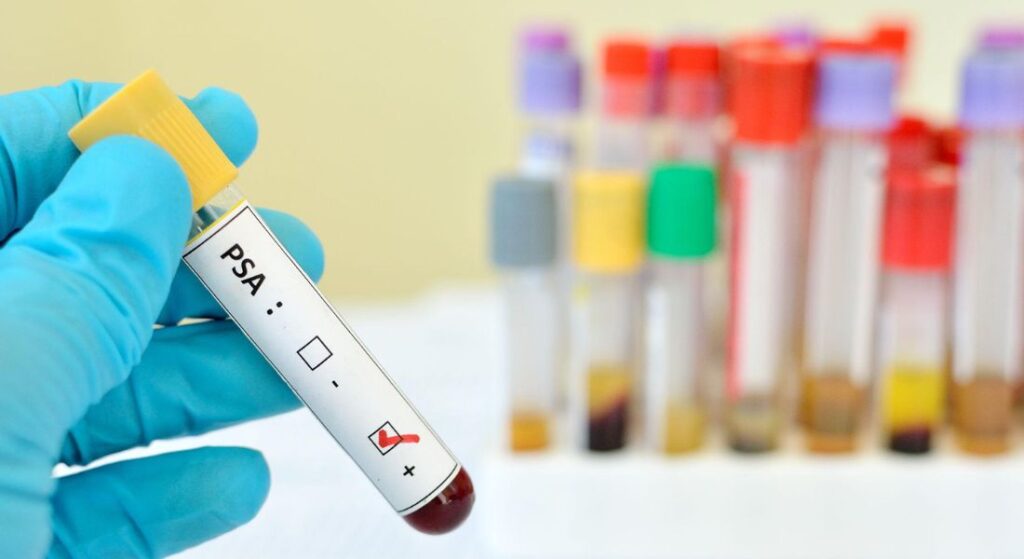
MRI provides detailed images of the prostate, but it does not replace the importance of your PSA blood test. Your PSA level, and how it changes over time, still gives valuable information about your prostate health. This helps your specialist monitor trends rather than relying on a single result.
A reassuring MRI combined with stable PSA levels and other normal findings may lead to a period of monitoring. However, if your PSA is rising or shows an unusual pattern, it may prompt closer investigation even if the MRI appears low risk. This ensures that subtle changes are not missed.
The relationship between your PSA results and MRI findings is more informative than either test on its own. Your specialist will interpret both together to build a clearer understanding of your situation. This combined approach helps guide the most appropriate plan for you.
Does a PI-RADS Score Tell You Whether You Need a Biopsy?
A PI-RADS score can strongly influence whether a biopsy is recommended, but it is not the only factor your specialist will consider. Higher scores are more likely to lead to a discussion about biopsy, while lower scores may support monitoring in suitable cases. This helps ensure that decisions are based on your overall risk, not just a single result.
In clinical practice, lesions scored from PI-RADS 3 to 5 are often reviewed more closely, with biopsy decisions guided by the wider clinical picture. Your specialist will assess how the MRI findings fit with other important information. This may include your PSA level, PSA density, age, symptoms, and previous biopsy history.
It is also important that your preferences are considered in the decision-making process. The choice about whether you need a biopsy should be explained clearly to you rather than being based only on a report number. This ensures you understand why a particular recommendation has been made for you.
What Is an MRI-Targeted Biopsy?
An MRI-targeted biopsy is where tissue samples are taken from areas your MRI has identified as suspicious. Using the imaging to guide the procedure allows your specialist to focus on the most relevant parts of your prostate, giving a more precise assessment.
- Targeting Suspicious Areas: The MRI highlights spots that may need closer examination, helping your specialist sample the areas most likely to be abnormal.
- Additional Samples: Depending on your situation, extra samples may be taken from other parts of the prostate to make sure nothing significant is missed.
- Confirming a Diagnosis: While the MRI shows how an area looks, only a biopsy can tell if cancer is present.
- Pathology Review: A pathologist examines the tissue in detail to provide a definitive diagnosis and guide your next steps.
Overall, an MRI-targeted biopsy helps give a clear and accurate picture of your prostate health. It allows your specialist to focus on the areas that matter most while ensuring a thorough assessment.
Can a Biopsy Be Negative After a High PI-RADS Score?
Yes, it is possible for a biopsy to be negative even if your MRI shows a high PI-RADS score. A PI-RADS score reflects how suspicious an area looks on imaging, but it does not confirm that cancer is definitely present. Not all suspicious-appearing areas turn out to be cancer.
If this happens, your specialist will review whether the biopsy adequately sampled the area seen on MRI. They will also consider whether there is still any ongoing concern based on your overall clinical picture. In some cases, further assessment such as monitoring, repeat imaging, or additional biopsy may be recommended.
A negative biopsy should always be interpreted in context rather than in isolation. You should receive a clear explanation of whether follow-up is needed and what changes would prompt further investigation. This helps ensure your care remains safe, structured, and tailored to you.
Can Cancer Be Found With a Low PI-RADS Score?

A low PI-RADS score reduces the likelihood of clinically significant prostate cancer, but it does not completely rule it out. MRI is a highly effective diagnostic tool, but no test can detect every single case of cancer with absolute certainty. This is why results always need to be interpreted carefully.
Specialists do not rely on MRI findings alone when assessing your risk. They also consider factors such as PSA density, PSA trends over time, examination findings, family history, and any previous test results. All of this information helps build a more complete picture of your prostate health.
A low PI-RADS score can be reassuring for you, but it is still important to follow the monitoring plan recommended by your specialist. Attending follow-up PSA tests and appointments ensures that any changes are detected early. This helps keep your care safe and appropriately managed over time.
Why Different Radiologists May Interpret MRI Differently
Prostate MRI interpretation requires specialist training and experience. Although PI-RADS provides a structured system for reporting, there can still be some variation in how borderline or complex findings are interpreted. This means that two experienced radiologists may occasionally describe subtle findings in slightly different ways.
Several factors can influence how an MRI is read, including image quality, scanner technique, and even patient movement during the scan. The level of radiologist experience can also play a role in interpretation. This is why high-quality imaging and specialist reporting are important for you.
In more complex cases, your clinical team may review your imaging in a multidisciplinary meeting or seek a second specialist opinion. This is particularly useful when MRI results do not fully match PSA levels or biopsy findings. It helps ensure that your diagnosis and treatment plan are as accurate and well-informed as possible for you.
PI-RADS and Likert Scores in UK Practice
You may receive either a PI-RADS score or a Likert score after prostate MRI, depending on the reporting system used by the imaging centre. Both use a five-point scale to communicate increasing levels of suspicion, but they are not identical scoring systems.
NICE guidance specifically recommends a five-point Likert scale within the UK prostate cancer diagnostic pathway and recommends MRI-influenced biopsy for a Likert score of 3 or more. PI-RADS, by comparison, uses structured rules based on MRI appearance, lesion location and the contribution of different MRI sequences.
For you, the practical message is that a PI-RADS 3 result and a Likert 3 result should not automatically be treated as technically identical simply because they use the same number. Your specialist should interpret the score according to the system used and combine it with your wider clinical risk.
Choosing Specialist Prostate MRI Assessment
Prostate MRI is most useful when high-quality imaging is combined with experienced interpretation and clear clinical follow-up. Your report should be assessed alongside your PSA level, prostate size, symptoms, and any previous test results. This helps ensure that the findings are meaningful for you rather than being viewed in isolation.
If you are considering specialist prostate MRI assessment, it is helpful to ask how your MRI findings will be reviewed and how PI-RADS results will guide decisions about biopsy or monitoring. A clear diagnostic pathway can reduce uncertainty and help you understand what each result means for you. This ensures you are not left with unanswered questions after your scan.
You should also feel comfortable asking who will explain your report to you in detail. Understanding a PI-RADS score becomes much easier when it is discussed in the context of your own prostate health. This personalised explanation helps you feel more informed and confident about your next steps.
Preparing for the Next Step After Your Result

The most important thing after receiving a PI-RADS score is to understand your individual next step. A low score may lead to monitoring, while an intermediate score may require a more personalised risk assessment. A higher score may lead to a discussion about biopsy to investigate the area further in your case.
It is important for you not to compare your result too closely with another patient’s experience. Differences in PSA level, prostate size, lesion features, age, family history, and previous test results can all influence the recommendation you receive. This means that the same score can lead to different plans for different people.
A PI-RADS score is designed to support clinical decision-making rather than provide a complete diagnosis on its own. The clearest approach is for you to discuss how your MRI result fits with your overall clinical picture. Following the investigation or monitoring plan recommended by your specialist will help ensure your care is appropriate and well-guided for you.
Myth vs Fact:
| Myth | Fact |
| PI-RADS 5 means stage 5 cancer. | PI-RADS is an MRI suspicion score, not a cancer stage. |
| PI-RADS 1 means cancer is impossible. | Low scores reduce concern but do not reduce the possibility of clinically significant cancer to zero. |
| PI-RADS 3 always means you need a biopsy. | It is an equivocal category, and decisions may consider PSA density and other clinical factors. |
| PI-RADS 4 confirms prostate cancer. | It indicates high imaging suspicion, but tissue diagnosis is still normally needed. |
| A negative biopsy always means the MRI was wrong. | The specialist may review targeting accuracy, pathology and ongoing clinical risk before deciding on further follow-up. |
| PI-RADS and Gleason scores are interchangeable. | PI-RADS describes MRI suspicion; Gleason grading and Grade Groups come from tissue examination. |
Key Takeaways:
- PI-RADS is a five-category MRI reporting system used to describe increasing levels of suspicion for clinically significant prostate cancer.
- A PI-RADS score is not a cancer diagnosis and should not be confused with cancer stage, Gleason score or Grade Group.
- PI-RADS 1 and 2 are low-suspicion categories, while PI-RADS 4 and 5 indicate progressively higher imaging concern.
- PI-RADS 3 is an equivocal category and often requires closer interpretation using PSA density and other clinical risk factors.
- Pooled research shows substantially higher cancer detection rates as scores rise from PI-RADS 3 to 5, although the exact probability varies between patient groups and centres.
- A high PI-RADS score still requires appropriate clinical assessment and usually tissue diagnosis before prostate cancer can be confirmed.
- PSA level, PSA density, prostate volume, family history, previous biopsy results and other findings can influence the next step.
- PI-RADS and Likert scoring both use five points but are separate systems; NICE guidance specifically frames the UK diagnostic pathway around Likert scoring.
FAQs:
1. Does a PI-RADS score mean that you have prostate cancer?
No, A PI-RADS score describes how suspicious an area looks on your prostate MRI, but it does not confirm that cancer is present. Higher scores are associated with greater concern for clinically significant prostate cancer, while lower scores are generally more reassuring. A biopsy may still be needed to confirm a diagnosis.
2. What is the difference between PI-RADS 3 and PI-RADS 4?
A PI-RADS 3 result is considered equivocal, which means the MRI finding is uncertain and cannot be clearly classified as either low or high suspicion. A PI-RADS 4 result means clinically significant prostate cancer is considered likely based on the MRI appearance. Decisions after a PI-RADS 3 result often depend more heavily on PSA density and other risk factors, while a PI-RADS 4 result commonly leads to discussion about biopsy.
3. Does a PI-RADS 5 score definitely mean prostate cancer?
No, PI-RADS 5 is the highest suspicion category, but it is still an imaging assessment rather than a confirmed diagnosis. Some non-cancerous conditions can occasionally produce suspicious MRI appearances. A biopsy is usually needed to determine whether cancer is present and, if so, what Grade Group it belongs to.
4. Can a biopsy be negative after a PI-RADS 4 or 5 result?
Yes, A biopsy can be negative even when the MRI shows a highly suspicious lesion. In this situation, your specialist may review whether the suspicious area was adequately sampled and whether the MRI, biopsy findings and PSA results agree with one another. Depending on your overall risk, follow-up may include PSA monitoring, specialist MRI review, repeat imaging or another biopsy.
5. Can clinically significant prostate cancer occur with a PI-RADS 1 or 2 score?
Yes, although the likelihood is lower. A low PI-RADS score is reassuring, but prostate MRI cannot detect every clinically significant cancer. Your specialist will therefore consider the MRI together with your PSA level, PSA density, family history, examination findings and previous test results. Continued monitoring may still be recommended even when the MRI result is low suspicion.
6. How does PSA density affect the interpretation of a PI-RADS 3 result?
PSA density compares your PSA level with the volume of your prostate. It can be particularly useful when a PI-RADS 3 lesion is found because the MRI result itself is uncertain. A specialist may use PSA density together with your PSA trend, age, family history and previous biopsy results when deciding whether biopsy or monitoring is more appropriate.
7. Is a PI-RADS score the same as a Gleason score or Grade Group?
No, PI-RADS is based on the appearance of your prostate on MRI and indicates the level of imaging suspicion. Gleason grading and Grade Groups are based on examining prostate tissue under a microscope after cancer has been confirmed through biopsy or surgery. You cannot directly convert a PI-RADS score into a Gleason score or Grade Group.
8. Does PI-RADS 5 mean stage 5 prostate cancer?
No, PI-RADS is not a cancer staging system, and a PI-RADS 5 result does not mean stage 5 cancer. The score only describes how suspicious a lesion appears on MRI. If cancer is confirmed, its stage is assessed separately using information such as biopsy results, examination findings and appropriate imaging.
9. What is the difference between a PI-RADS score and a Likert score?
Both PI-RADS and Likert scoring use five categories to describe the level of suspicion seen on prostate MRI, but they are separate reporting systems. PI-RADS uses structured MRI interpretation criteria, while Likert scoring relies more on the radiologist’s overall assessment of the scan. Your specialist should interpret the score according to the system used by the imaging centre and combine it with your wider clinical information.
10. Can your PI-RADS score change on a future MRI?
Yes, A lesion may appear different on a future MRI because of genuine biological change, changes in inflammation or benign prostate conditions, differences in scan quality, or differences in interpretation. A change in PI-RADS score should be reviewed alongside your PSA trend, PSA density, previous biopsy findings and other clinical information. Your specialist can then decide whether continued monitoring, repeat imaging or biopsy is appropriate.
Final Thoughts: Understanding Your PI-RADS Score in Context
Receiving a PI-RADS score can understandably raise questions, but it is important to remember that the score is only one part of a much larger clinical assessment. While the number helps indicate how suspicious an area appears on MRI, it does not confirm whether cancer is present and should never be interpreted in isolation. Your PSA level, PSA density, prostate size, medical history, family history, examination findings, and any biopsy results all contribute to understanding your individual level of risk.
The most valuable next step is discussing your MRI findings with a specialist who can explain what the score means for you personally. Whether your result leads to monitoring, further imaging, or a biopsy, having a clear plan can help reduce uncertainty and ensure that any necessary investigations are carried out appropriately.
If you are considering assessment with a specialist team offering a prostate MRI scan in London, you can contact us to discuss your options and arrange a consultation tailored to your individual needs.
References:
- National Institute for Health and Care Excellence (NICE) (2019, reviewed 2025) Prostate cancer: diagnosis and management. NICE guideline NG131. Available at: https://www.nice.org.uk/guidance/ng131
- Turkbey, B. et al. (2019) ‘Prostate Imaging Reporting and Data System Version 2.1: 2019 Update of Prostate Imaging Reporting and Data System Version 2’, European Urology, 76(3), pp.340–351. Available at: https://pubmed.ncbi.nlm.nih.gov/30898406/
- Oerther, B. et al. (2024) ‘Update on PI-RADS Version 2.1 Diagnostic Performance Benchmarks for Prostate MRI: Systematic Review and Meta-Analysis’, Radiology, 312(2), e233337. Available at: https://pubmed.ncbi.nlm.nih.gov/39136561/
- European Association of Urology (EAU) (2026) EAU Guidelines on Prostate Cancer: Diagnostic Evaluation. Available at: https://uroweb.org/guidelines/prostate-cancer/chapter/diagnostic-evaluation
- Kasivisvanathan, V. et al. (2018) ‘MRI-Targeted or Standard Biopsy for Prostate-Cancer Diagnosis’, New England Journal of Medicine, 378(19), pp.1767–1777. Available at: https://pubmed.ncbi.nlm.nih.gov/29552975/