




For many years, diagnosing prostate cancer relied mainly on systematic biopsies, where multiple samples were taken from different areas of the prostate using ultrasound guidance. While this approach helped detect cancer, it could sometimes miss important tumours or identify lower-risk cancers that may never have caused problems during your lifetime.
The PRECISION Trial changed how specialists approach prostate biopsy by demonstrating the value of using MRI before deciding whether a biopsy is needed. If you are being assessed for possible prostate cancer, an MRI-based pathway can provide more detailed information about suspicious areas and help doctors identify cancers that are more likely to require treatment.
The study showed that MRI-guided assessment could improve the detection of clinically significant prostate cancers while reducing the diagnosis of some lower-risk cancers. Today, MRI plays an important role in many prostate cancer diagnostic pathways, helping your specialist make more informed decisions about whether you need a biopsy and where samples should be taken.
What Was the PRECISION Trial?
The PRECISION Trial was a major international study that compared two different approaches to diagnosing prostate cancer. If you are being investigated for possible prostate cancer, the findings from this trial can help you understand why MRI has become such an important part of modern diagnostic pathways.
The study compared traditional ultrasound-guided systematic biopsy with an MRI-based approach followed by targeted biopsy when suspicious areas were found. If you were part of the MRI group, the scan helped doctors identify areas that required closer assessment rather than taking samples randomly from across the prostate.
The trial included men with a clinical suspicion of prostate cancer who had not previously undergone a prostate biopsy. The aim was to determine whether MRI could improve diagnostic accuracy and reduce unnecessary procedures. Today, your specialist may use this evidence when deciding whether you need a biopsy and which areas of your prostate should be targeted.
Why Was the Trial Needed?
Before MRI became widely used in prostate cancer assessment, many men with raised PSA levels were referred directly for biopsy. If you had an elevated PSA result, you may have gone straight to a traditional biopsy where samples were taken from different areas of the prostate without knowing exactly where significant cancer might be present.
This approach created two important challenges. Some cancers that needed treatment could be missed, while you could also be diagnosed with small, low-risk cancers that may never have caused problems. This sometimes led to unnecessary worry, further investigations, and difficult treatment decisions.
The PRECISION Trial explored whether MRI could make the diagnostic process more accurate and personalised. By providing clearer information about your prostate, MRI helps your specialist decide whether a biopsy is needed and identify the areas most likely to contain clinically significant cancer.
How Were Prostate Biopsies Traditionally Performed?
Traditional prostate biopsy commonly involved taking multiple tissue samples using transrectal ultrasound guidance. If you were undergoing this type of biopsy, the ultrasound helped guide the needle placement, but it could not reliably show every area where prostate cancer might be present.
Because suspicious areas were not always visible, doctors took samples systematically from different regions of the prostate gland. This approach helped detect many cancers, but it relied on the possibility that the biopsy needle would pass through an area containing significant disease.
Although this method played an important role in prostate cancer diagnosis, it was considered a less targeted approach. When you had a systematic biopsy, samples were not always taken from a specific abnormal area identified through imaging, which meant some important cancers could potentially be missed.
What Is an MRI-Targeted Biopsy?
An MRI-targeted biopsy uses information from a prostate MRI scan to identify areas that appear suspicious. If you are being assessed for possible prostate cancer, this approach allows your specialist to see which parts of the prostate may need closer examination before taking biopsy samples.
Instead of relying only on systematic sampling, the biopsy can be directed towards specific areas that look abnormal on the MRI scan. This means doctors can focus on regions that are more likely to contain clinically significant cancer, helping to make the diagnostic process more precise.
The PRECISION Trial showed that using MRI before biopsy could improve the detection of important cancers compared with traditional biopsy pathways. For you, this means modern diagnostic approaches may provide clearer information about whether cancer is present and whether further treatment decisions are needed.
What Did the PRECISION Trial Find?
One of the most important findings from the PRECISION Trial was that an MRI-based pathway detected more clinically significant prostate cancers compared with standard ultrasound-guided biopsy. If you are being investigated for prostate cancer, this means MRI can provide additional information to help identify cancers that are more likely to require attention.
The trial also showed that some men with a low-risk MRI result could safely avoid an immediate biopsy. This approach may help you avoid an unnecessary procedure while still allowing your specialist to monitor your situation and recommend further investigation if needed.
The findings changed how doctors think about prostate biopsy. The goal is no longer simply to find any cancer, but to identify cancers that are more likely to affect your health and require treatment. This helps your specialist make more personalised decisions based on your individual risk rather than relying only on traditional biopsy methods.
Evidence Note:
The PRECISION Trial showed that an MRI-based pathway changed prostate cancer diagnosis in several important ways. In the MRI group, 71 of 252 men, or 28%, avoided biopsy because their MRI did not show suspicious areas. Clinically significant prostate cancer was detected in 38% of men in the MRI-targeted pathway compared with 26% in the standard biopsy pathway. The MRI pathway also reduced detection of clinically insignificant prostate cancer, reported as 9% compared with 22% in the standard biopsy group. For you as a patient, this means MRI can help doctors focus biopsy on cancers that are more likely to matter clinically, while reducing unnecessary procedures and some low-risk diagnoses.
How Did MRI Change the Diagnostic Process?
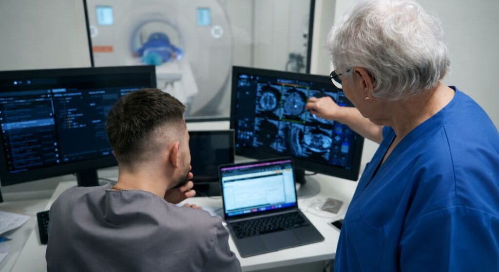
Before the PRECISION Trial, many men followed a pathway where a raised PSA level often led directly to a prostate biopsy. If you had an abnormal PSA result, the next step was frequently an invasive test, even though it was not always clear whether significant cancer was present.
MRI introduced an additional decision-making stage between suspicion and biopsy. Your specialist can now use MRI findings to assess whether a biopsy is likely to be beneficial and identify specific areas of the prostate that may need targeted sampling.
This has made prostate cancer diagnosis more selective and personalised. Instead of following the same pathway for every patient, doctors can use your MRI results, PSA level, and other risk factors to decide the most appropriate next step for your individual situation.
Why Detecting Clinically Significant Cancer Matters
Not all prostate cancers behave in the same way. Some cancers grow very slowly and may never cause symptoms or affect your health, while others have features that make them more likely to grow, spread, and require treatment.
The aim of modern prostate cancer diagnosis is not simply to find any cancer cells, but to identify cancers that are clinically significant. If you are being assessed for prostate cancer, this approach helps your specialist focus on finding disease that is more likely to affect you while avoiding unnecessary investigations and treatment where possible.
The PRECISION Trial helped support this change in thinking by showing that MRI-targeted pathways could improve the detection of important cancers while reducing some unnecessary diagnoses. This means your diagnostic pathway can be more personalised, using imaging and other information to guide decisions that are more specific to your individual risk.
Did the Trial Mean Fewer Men Needed Biopsies?
One key finding from the PRECISION Trial was that some men with MRI scans showing no suspicious areas could safely avoid an immediate biopsy. This approach helps reduce the number of invasive procedures when the chance of detecting significant cancer is low.
- Reducing Unnecessary Biopsies: Using MRI first allows specialists to focus on men more likely to benefit from a biopsy, sparing others from unnecessary procedures.
- Maintaining Focus on Significant Disease: The approach prioritises detecting clinically important cancers while avoiding low-risk overdiagnosis.
- MRI Complements Clinical Judgement: Decisions are based on PSA, family history, examination, MRI findings, and other risk factors, not the scan alone.
- Individualised Decision Making: Your doctor will use MRI information alongside other clinical data to decide the most appropriate next steps for you.
Overall, the PRECISION Trial showed that MRI can help some men avoid unnecessary biopsies, but careful assessment and personalised clinical judgement remain essential.
Does MRI Replace Prostate Biopsy Completely?
No. MRI has significantly improved the way prostate cancer is diagnosed, but it does not completely replace the need for a biopsy. If you are being assessed for possible prostate cancer, MRI is an important tool, but it is usually used alongside other investigations rather than as a standalone test.
A biopsy may still be recommended when your MRI shows suspicious areas or when other factors suggest that cancer could be present. Your specialist will consider your PSA level, MRI findings, symptoms, and overall risk profile before deciding whether tissue sampling is needed.
MRI helps doctors identify who is more likely to benefit from a biopsy and where samples should be taken. However, tissue analysis remains essential in many cases because it provides detailed information about the type and grade of cancer, helping you and your healthcare team make informed treatment decisions.
Clinical Tip: MRI Guides Biopsy, But Tissue Still Matters
A prostate MRI can show areas that look suspicious, but it cannot confirm cancer by itself. A biopsy is still needed when tissue diagnosis will change your care, because the laboratory report provides the cancer grade, pattern and other details that guide treatment decisions. A reassuring MRI can reduce the likelihood of clinically significant cancer, but your specialist will still consider PSA density, family history, symptoms and other risk factors before deciding whether biopsy can safely be avoided.
What Is a PI-RADS Score?
After a prostate MRI, the radiologist may assign a PI-RADS score to describe how suspicious an area of the prostate appears for clinically significant prostate cancer. If you are having an MRI as part of your assessment, this score can help your specialist understand the level of concern and decide what steps may be needed next.
The PI-RADS system uses a scale to indicate the likelihood that an abnormal area may contain significant cancer. Higher scores generally suggest a greater level of suspicion, while lower scores indicate that clinically significant cancer is less likely.
Your PI-RADS score is only one part of the overall assessment. Your specialist will also consider your PSA level, biopsy results if available, family history, and other risk factors before recommending whether you need a biopsy, further monitoring, or another investigation.
PI-RADS Score Interpretation and Recommended Biopsy Approach
| PI-RADS score | Likelihood of clinically significant cancer | Typical next step | Notes |
| 1 | Very low | Monitoring is often considered | MRI appears normal, but PSA and risk factors still matter |
| 2 | Low | Monitoring or repeat assessment may be appropriate | Biopsy is often not needed immediately unless other risks are concerning |
| 3 | Intermediate / equivocal | Biopsy may be considered | Decision depends on PSA density, age, family history and overall risk |
| 4 | High | Targeted biopsy is commonly recommended | Suspicious area is more likely to contain significant cancer |
| 5 | Very high | Targeted biopsy is usually strongly considered | Prompt specialist evaluation is usually appropriate |
How Did PRECISION Reduce Overdiagnosis?
One of the long-standing challenges in prostate cancer diagnosis has been identifying which cancers are likely to cause harm and which may remain slow-growing without affecting your health. Traditional biopsy methods could sometimes detect small, low-risk cancers that may not have required immediate treatment.
If you were diagnosed with a low-risk cancer, this could create difficult decisions about whether to undergo treatment or continue monitoring. The PRECISION Trial helped support a more targeted approach by showing that MRI-based pathways could improve the identification of cancers that are more likely to be clinically significant.
By focusing on cancers that are more likely to affect you, MRI-targeted diagnosis aims to reduce unnecessary diagnoses and treatments while still detecting important disease. This allows your specialist to provide a more personalised assessment based on the likelihood that the cancer may require intervention.
Why Was the Trial Considered Practice-Changing?
The PRECISION Trial was considered an important study because it provided strong evidence from a randomised clinical trial rather than relying only on observational research. If you are being assessed for possible prostate cancer, the findings help explain why your diagnostic pathway may now begin with MRI before deciding whether a biopsy is needed.
The results supported a move towards MRI-first approaches, where imaging is used to provide more information before invasive testing takes place. This can help your specialist identify suspicious areas more accurately and decide whether a targeted biopsy is appropriate.
The trial influenced discussions around prostate cancer guidelines and changed expectations about how men with suspected prostate cancer should be assessed. Today, MRI plays a much larger role in diagnosis, helping you receive a more personalised evaluation based on your individual risk factors and test results.
How Did the Trial Affect Patient Experience?

For many men, being told that a prostate biopsy is needed can create anxiety and uncertainty. If you are facing this situation, knowing that MRI can help decide whether a biopsy is necessary may provide reassurance and reduce the chance of undergoing an avoidable procedure.
MRI also gives you and your specialist more information about what is happening inside the prostate before further investigations are considered. By identifying areas that may need closer attention, it can make the diagnostic process feel more focused and easier to understand.
A clearer pathway can help you become more involved in decisions about your care. When you understand why a biopsy is recommended or why monitoring may be suitable, you can feel more confident about the next steps in your prostate cancer assessment.
What Are the Benefits of MRI Before Biopsy?
MRI provides detailed images of the prostate and surrounding tissues, helping your specialist identify areas that may require closer investigation. If you are being assessed for possible prostate cancer, this additional information can provide a clearer picture of what is happening inside the prostate before deciding on further tests.
Instead of relying only on systematic sampling, MRI allows biopsy samples to be directed towards areas that appear suspicious. This targeted approach means doctors can focus on regions that are more likely to contain clinically significant cancer rather than taking samples without specific guidance.
Using MRI before biopsy can improve diagnostic accuracy and help reduce unnecessary procedures for selected patients. By combining your MRI findings with PSA levels and other clinical information, your healthcare team can create a more personalised diagnostic plan.
What Happens If MRI Shows a Suspicious Area?
If your MRI identifies an area that appears suspicious, your specialist may recommend an MRI-targeted biopsy to investigate it further. This allows the biopsy to focus on the specific region that requires assessment rather than relying only on samples taken from different areas of the prostate.
During the procedure, your doctor uses the information from your MRI scan to guide the biopsy towards the area of concern. This targeted approach can help improve the chances of detecting clinically significant cancer if it is present.
The exact biopsy method recommended for you will depend on several factors, including your healthcare provider, available technology, MRI findings, and individual circumstances. Your specialist can explain which approach is most suitable and what you can expect during the procedure.
Understanding the Role of Targeted Biopsy
Targeted biopsy has changed the focus of prostate cancer diagnosis from simply taking more samples to taking more meaningful samples. If you are being assessed for possible prostate cancer, this approach allows your specialist to use detailed MRI information to decide which areas of the prostate need closer investigation.
Instead of relying only on samples taken from random areas of the gland, doctors can use MRI findings to guide where suspicious tissue should be collected. This helps create a more focused diagnostic process and may improve the likelihood of identifying important cancers.
The aim of targeted biopsy is to improve the detection of clinically significant prostate cancer while reducing the unnecessary identification of very low-risk disease. By using a more personalised approach, you and your healthcare team can make better-informed decisions about whether further treatment or monitoring is appropriate.
Clinical Nuance:
In the PRECISION Trial, men with suspicious MRI findings in the MRI pathway underwent targeted biopsy. In current practice, some specialists may recommend targeted biopsy alone, while others may combine targeted and systematic samples depending on MRI findings, PSA density, prostate size, previous biopsy history and overall risk. Your specialist should explain which approach is being recommended and why.
What Happens During an MRI-Targeted Biopsy?
Before an MRI-targeted biopsy, you will usually have a prostate MRI scan to identify areas that may require further assessment. If your scan shows a suspicious area, your specialist can use this information to plan where biopsy samples should be taken.
During the procedure, MRI findings are combined with ultrasound guidance or specialised fusion technology to help your doctor target the area of concern more accurately. This allows the biopsy to focus on specific regions rather than relying only on a standard systematic approach.
The procedure still involves taking small tissue samples from the prostate, which are then examined by a specialist pathologist. The results provide important information about whether cancer is present, how aggressive it may be, and what treatment options may be suitable for you.
Biopsy Safety Note:
Temporary blood in the urine or semen, bruising and discomfort can occur after prostate biopsy. Less common complications include urinary retention, significant bleeding and infection. Transrectal biopsy can rarely lead to sepsis.
Seek urgent medical advice if you develop fever, chills, worsening pain, difficulty passing urine, heavy or persistent bleeding, confusion, faintness or feel acutely unwell after biopsy. Follow the specific emergency instructions given by the biopsy team.
Why Tissue Diagnosis Is Still Important

Although MRI provides valuable information about the prostate, it cannot confirm cancer on its own. If your MRI shows a suspicious area, a biopsy may still be needed to examine the tissue directly and provide a definite diagnosis.
A biopsy allows doctors to study prostate cells under a microscope and determine whether cancer is present and how aggressive it appears. This information helps your specialist understand the nature of the cancer rather than relying only on imaging results.
The biopsy report provides important details, including the cancer grade and other features that help guide treatment decisions. By combining your biopsy results with MRI findings, PSA levels, and other factors, your healthcare team can recommend an approach that is most appropriate for your situation.
How PRECISION Changed the PSA Pathway
Before MRI became widely integrated into prostate cancer diagnosis, many men with raised PSA levels were often referred directly for biopsy. If you had an elevated PSA result, you may have moved quickly towards an invasive test without having detailed information about whether a significant cancer was likely to be present.
The PRECISION Trial supported a different approach, where MRI is used as an additional step to help decide whether a biopsy is needed and how it should be performed. Your MRI results can help your specialist identify suspicious areas and determine whether targeted sampling may provide more useful information.
This has made the diagnostic pathway more focused and personalised. Instead of performing biopsies in the same way for every patient, doctors can use your PSA level, MRI findings, and other risk factors to identify those who are most likely to benefit from invasive testing.
Does Everyone with a Raised PSA Need an MRI?
A raised PSA level does not always mean that you have prostate cancer. PSA can increase for several reasons, including an enlarged prostate, inflammation, infection, or other non-cancerous conditions.
If you have an elevated PSA result, your specialist will consider several factors before recommending the next step. These may include your PSA level, age, symptoms, family history, examination findings, and other individual risk factors.
MRI is an important part of modern prostate assessment, but it is not automatically required for everyone with a raised PSA. Your specialist will decide whether an MRI is appropriate based on your overall situation and whether it is likely to provide useful information for your diagnosis and care.
Can a Normal MRI Rule Out Prostate Cancer?
A reassuring MRI result can significantly reduce the likelihood of clinically significant prostate cancer, but it does not completely rule out the possibility. If you have a normal MRI, your specialist will still consider the result alongside your other clinical information before deciding on the next step.
Factors such as your PSA level, family history, age, symptoms, and overall risk profile can influence whether further assessment is needed. Your doctor will look at the complete picture rather than relying on the MRI result alone.
In some situations, you may only need ongoing monitoring with regular PSA tests and reviews. However, if other risk factors suggest that cancer could still be present, your specialist may recommend additional investigations or follow-up to ensure any important changes are identified early.
How Has MRI Improved Shared Decision-Making?
Before MRI became more established in prostate cancer assessment, some men faced uncertainty about whether a biopsy was necessary. If you were in this situation, it could be difficult to understand whether an invasive test was likely to provide useful information or whether monitoring might be more appropriate.
MRI provides additional detail about the prostate, helping your specialist explain why a biopsy may or may not be recommended. By identifying suspicious areas or showing a lower likelihood of significant cancer, MRI can make the reasoning behind the next steps clearer.
This allows you to have a more informed discussion about the potential benefits, risks, and outcomes of further testing. Understanding why a particular approach is suggested can help you feel more involved in decisions about your diagnosis and care.
What Are the Limitations of MRI?
MRI has greatly improved prostate cancer diagnosis, but it isn’t a perfect test. While it provides valuable information, it cannot detect every cancer with complete certainty.
- Cancers May Be Missed: Some tumours might not be clearly visible on MRI, so a “normal” scan doesn’t guarantee the absence of cancer.
- False Suspicious Areas: Areas that look abnormal on MRI may not actually contain cancer, so results need careful interpretation.
- Combined Assessment Needed: Specialists consider MRI results alongside PSA levels, symptoms, family history, examination findings, and any biopsy results.
- Personalised Decision Making: Using all available information together gives a more accurate picture of your individual risk and the next steps in your care.
Overall, MRI is a powerful tool but must be interpreted in context. A combined assessment helps ensure your diagnosis and treatment plan are tailored to your specific situation.
How Did PRECISION Influence Modern Guidelines?
The PRECISION Trial contributed to a major change in how prostate cancer diagnosis is approached. If you are being assessed for suspected prostate cancer today, your diagnostic pathway may include multiparametric MRI before deciding whether a biopsy is needed.
Many professional guidelines now recommend considering MRI before biopsy because it can provide important information about areas of concern within the prostate. This helps your specialist decide whether a biopsy is appropriate and whether targeted sampling may be more beneficial than traditional approaches alone.
The trial helped establish MRI as an important tool for improving diagnostic accuracy and reducing unnecessary procedures. By incorporating MRI into the assessment process, you can benefit from a more personalised pathway that considers your individual risk factors and clinical findings.
UK Guidance Note:
In UK practice, NICE recommends multiparametric MRI as the first-line investigation for people with suspected clinically localised prostate cancer. NICE also recommends offering MRI-influenced prostate biopsy when the MRI score is 3 or more, and considering whether biopsy can be omitted when the MRI score is low, but only after discussing the risks and benefits with the patient. This means MRI has become a key step in the diagnostic pathway, but biopsy decisions should still be personalised using PSA, examination findings, MRI results, risk factors and shared decision-making.
What Does This Mean If You Need a Prostate Biopsy?
If your doctor recommends a prostate biopsy, it does not necessarily mean that cancer has already been confirmed. If you are advised to have a biopsy, it usually means that your PSA results, MRI findings, or other clinical information suggest that more detailed assessment is needed.
A biopsy is used to examine prostate tissue directly and provide a clearer understanding of what is happening inside the gland. Your specialist can use the results to determine whether cancer is present and, if so, understand its characteristics.
MRI has helped make many biopsies more targeted by showing areas that may require closer investigation. This means your doctor can use imaging information to guide sampling and gain more accurate information about any abnormal areas within the prostate.
How Has PRECISION Changed Patient Care?
The biggest change from the PRECISION Trial is that prostate cancer diagnosis has become more personalised. If you are being assessed for possible prostate cancer, you may now follow a pathway that uses MRI information to help decide the most appropriate next step rather than automatically moving straight to biopsy.
Instead of every man following the same diagnostic process, doctors can use your MRI results, PSA level, and other risk factors to guide decisions. This allows your specialist to identify when a biopsy is more likely to be useful and when monitoring may be a suitable option.
This approach aims to provide the right investigation for the right patient at the right time. By making diagnosis more targeted, you can benefit from a clearer assessment process that reduces unnecessary procedures while improving the detection of cancers that are more likely to need treatment.
The Importance of Specialist MRI Interpretation

A prostate MRI is a detailed investigation that requires careful interpretation by experienced specialists. If you are having an MRI as part of your prostate cancer assessment, the accuracy of the findings can depend on both the quality of the scan and the expertise of the radiologist reviewing the images.
The experience of the reporting specialist can influence how confidently suspicious areas are identified and classified. A well-interpreted MRI can provide valuable information to help guide decisions about whether a biopsy or further monitoring may be appropriate.
Your MRI result should always be considered alongside your complete clinical picture rather than viewed as an isolated report. Your specialist will also consider factors such as your PSA level, symptoms, examination findings, and other risk factors to provide a more accurate assessment of your individual situation.
How Does This Affect Prostate Biopsy Decisions?
The decision to perform a prostate biopsy depends on several factors rather than MRI results alone. If you are being assessed for possible prostate cancer, your specialist will look at the complete picture to decide whether a biopsy is likely to provide useful information.
Your MRI findings may be considered alongside your PSA level, PSA density, family history, previous test results, and personal preferences. This means the recommendation for a biopsy is tailored to your individual risk rather than based on a single test result.
If you are considering assessment through a specialist prostate biopsy in London, your consultation should include a clear explanation of why a biopsy is being recommended and what information it can provide. Understanding the purpose of the procedure can help you feel more confident when making decisions about your diagnosis and care.
Moving Forward After the PRECISION Trial
The PRECISION Trial changed prostate cancer diagnosis by showing that MRI can play a central role before deciding whether a biopsy is needed. If you are being assessed for possible prostate cancer today, your diagnostic pathway may include MRI to provide more information before moving forward with invasive testing.
The study helped move care away from a purely biopsy-first approach and towards a more informed imaging-based pathway. By using MRI findings alongside PSA levels and other risk factors, your specialist can make a more personalised decision about whether a biopsy is appropriate.
The main lesson from the trial is that prostate cancer diagnosis is not only about finding any cancer. It is about identifying cancers that are more likely to affect your health while reducing unnecessary procedures where possible. This approach helps ensure that you receive the right investigation at the right time based on your individual circumstances.
Myth vs Fact:
| Myth | Fact |
| MRI means you will never need a prostate biopsy. | MRI can reduce unnecessary biopsies, but biopsy is still needed when tissue diagnosis is important. |
| A normal MRI rules out prostate cancer completely. | A reassuring MRI lowers the chance of significant cancer but does not remove the risk completely. |
| A raised PSA always means cancer. | PSA can rise due to BPH, inflammation, infection and other non-cancerous causes. |
| Targeted biopsy finds every important cancer. | Targeting improves accuracy, but no test is perfect. |
| Systematic biopsy is now useless. | Systematic sampling may still be used in selected cases, sometimes alongside targeted biopsy. |
| PI-RADS or Likert score alone decides everything. | MRI score is interpreted alongside PSA, PSA density, examination findings and personal risk factors. |
Key Takeaways:
- The PRECISION Trial showed that MRI before biopsy can improve prostate cancer diagnosis.
- In the MRI pathway, 28% of men in the MRI-pathway group avoided an immediate biopsy because the MRI was not suspicious.
- MRI-targeted biopsy detected more clinically significant prostate cancer than standard biopsy in the trial.
- The MRI pathway also reduced diagnosis of some clinically insignificant cancers.
- MRI does not completely replace biopsy because tissue diagnosis is still needed in many cases.
- A normal MRI lowers the chance of significant cancer but does not rule it out completely.
- Modern biopsy decisions should consider MRI results, PSA, PSA density, examination findings, family history and personal risk factors.
FAQs:
1. What was the main finding of the PRECISION Trial?
The PRECISION Trial showed that using MRI before prostate biopsy improved detection of clinically significant prostate cancer while reducing diagnoses of clinically insignificant cancer. It demonstrated that an MRI-based pathway could help identify men who were more likely to benefit from a targeted biopsy. The study helped change prostate cancer diagnosis by making the process more accurate and personalised.
2. Why was the PRECISION Trial important for prostate cancer diagnosis?
Before the trial, many men with a raised PSA level were referred directly for a systematic biopsy. The PRECISION Trial showed that MRI could provide additional information before biopsy and help doctors make better decisions. This reduced unnecessary procedures while improving the detection of important cancers.
3. Does the PRECISION Trial mean you can avoid a prostate biopsy?
In some cases, a low-suspicion MRI may support a shared decision to omit or defer an immediate biopsy when other risk factors are also reassuring. However, MRI does not completely replace biopsy, as tissue samples are still required when there is a higher risk of significant cancer. Your specialist will consider your MRI findings, PSA level and other risk factors before recommending the next step.
4. How does an MRI-targeted biopsy differ from a traditional prostate biopsy?
A traditional biopsy involves taking samples from different areas of the prostate using ultrasound guidance according to a planned anatomical pattern rather than an MRI-visible target. An MRI-targeted biopsy uses scan images to guide samples towards areas that appear suspicious. This allows doctors to focus on areas that are more likely to contain clinically significant cancer.
5. What does a PI-RADS score mean after a prostate MRI?
A PI-RADS score shows how suspicious an area of the prostate appears on an MRI scan for clinically significant cancer. Higher scores generally indicate a greater likelihood that further investigation may be needed. Your specialist will consider your PI-RADS score alongside your PSA level, medical history and other risk factors.
6. Can a prostate MRI detect all types of prostate cancer?
No, a prostate MRI cannot detect every prostate cancer with complete certainty. Some cancers may not be clearly visible, while some suspicious areas may not contain cancer. Your specialist will combine your MRI results with other information to decide whether further investigation is needed.
7. Why is detecting clinically significant prostate cancer important?
Not all prostate cancers behave in the same way, and some slow-growing cancers may never cause health problems. The aim of modern diagnosis is to identify cancers that are more likely to grow or require treatment. The PRECISION Trial supported this approach by helping doctors focus on finding cancers that matter most.
8. Does everyone with a raised PSA level need a prostate MRI?
A raised PSA level does not always mean you have prostate cancer, as it can increase for several other reasons. Your specialist will consider your age, symptoms, PSA level, family history and other factors before deciding whether an MRI is appropriate. MRI is now an important part of assessment for many men with suspected prostate cancer.
9. What happens if your MRI shows a suspicious area?
If your MRI identifies an area of concern, your specialist may recommend a targeted biopsy to examine that area more closely. The biopsy samples are analysed to confirm whether cancer is present and understand its characteristics. This information helps guide decisions about monitoring or treatment.
10. How has the PRECISION Trial changed patient care?
The PRECISION Trial helped introduce a more personalised approach to prostate cancer diagnosis. Instead of automatically moving from a raised PSA result to biopsy, doctors can now use MRI findings to guide decisions. This allows you to receive the most appropriate investigation while reducing unnecessary procedures where possible.
Final Thoughts: How the PRECISION Trial Improved Prostate Cancer Diagnosis
The PRECISION Trial showed that an MRI-led diagnostic pathway could identify more clinically significant prostate cancers and fewer clinically insignificant cancers than standard systematic biopsy. It also allowed 28% of men allocated to the MRI pathway to avoid an immediate biopsy because their scan was not suspicious.
MRI is not a perfect test and does not remove the need for tissue diagnosis in every case. A low-suspicion scan can still miss clinically significant disease, while a suspicious scan does not confirm cancer. Biopsy decisions should therefore consider MRI findings alongside PSA density, examination findings, family history and your wider clinical risk.
If you are considering a prostate biopsy in London, you can contact us at Prostate Clinic London to discuss appointment arrangements, the reasons a biopsy may or may not be recommended and the biopsy approaches available.
References:
- National Institute for Health and Care Excellence (NICE) (2019, last updated 2021; reviewed 2025) Prostate cancer: diagnosis and management. NICE guideline NG131. Available at: https://www.nice.org.uk/guidance/ng131
- Kasivisvanathan, V., Rannikko, A.S., Borghi, M., Panebianco, V., Mynderse, L.A., Vaarala, M.H., Briganti, A., Budäus, L., Hellawell, G., Hindley, R.G. et al. (2018) ‘MRI-Targeted or Standard Biopsy for Prostate-Cancer Diagnosis’, New England Journal of Medicine, 378(19), pp.1767–1777. Available at: https://pubmed.ncbi.nlm.nih.gov/29552975/
- Ahmed, H.U., El-Shater Bosaily, A., Brown, L.C., Gabe, R., Kaplan, R., Parmar, M.K.B., Collaco-Moraes, Y., Ward, K., Hindley, R.G., Freeman, A. et al. (2017) ‘Diagnostic accuracy of multi-parametric MRI and TRUS biopsy in prostate cancer (PROMIS): a paired validating confirmatory study’, The Lancet, 389(10071), pp.815–822. Available at: https://pubmed.ncbi.nlm.nih.gov/28110982
- National Institute for Health and Care Excellence (2019, updated 2021, reviewed 2025) Prostate cancer: diagnosis and management (NG131). Available at: https://www.nice.org.uk/guidance/ng131
- Drost, F.J.H., Osses, D.F., Nieboer, D., Bangma, C.H., Steyerberg, E.W., Roobol, M.J. and Schoots, I.G. (2020) ‘Prostate magnetic resonance imaging, with or without magnetic resonance imaging-targeted biopsy, and systematic biopsy for detecting prostate cancer: a Cochrane systematic review and meta-analysis’, European Urology, 77(1), pp.78–94. Available at: https://pubmed.ncbi.nlm.nih.gov/31326219/
- Turkbey, B., Rosenkrantz, A.B., Haider, M.A., Padhani, A.R., Villeirs, G., Macura, K.J., Tempany, C.M., Choyke, P.L., Cornud, F., Margolis, D.J.A. et al. (2019) ‘Prostate Imaging Reporting and Data System Version 2.1: 2019 update of Prostate Imaging Reporting and Data System Version 2’, European Urology, 76(3), pp.340–351. Available at: https://pubmed.ncbi.nlm.nih.gov/30898406/